 Find a Doctor
  Locations
  Services
 McLeod Performs First Brain Coiling Procedure
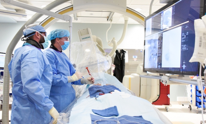
Photo: McLeod leads the way in technology with the performance of the area’s first procedure for an unruptured brain aneurysm in an operating suite dedicated for this work. Interventional Radiologist Dr. Chris Durst led the Neurovascular Team and is pictured here with Specials Procedures Radiology Technologist Shayne Anderson.
An Advanced Procedure to Reduce Stroke Risk
Using only a tiny metal spring, the medical experts at McLeod lowered a patient’s risk of stroke when they performed the area’s first advanced procedure for an unruptured brain aneurysm in an operating suite dedicated for this work.
The McLeod Neurovascular Team completed their first coiling procedure for an unruptured brain aneurysm. The coiling, executed by Interventional Radiologist Dr. Chris Durst, was performed in the new dedicated Neurointerventional Bi-plane Suite at McLeod Regional Medical Center.
“This is an exciting time for our Neurovascular Team,” said Dr. Timothy Hagen, McLeod Medical Director, Stroke and Neurology Services. “Brain aneurysms are a serious medical condition. If they rupture it could cause a serious stroke or even death. This technique can prevent rupturing and saves lives. We are glad to be able to offer this amazing life-saving procedure for our patients.”
A brain aneurysm is a bulging area within the wall of an artery that supplies blood and oxygen to the brain. As the blood pressure in the artery pushes on this protruding spot the danger is that it can continue to balloon out with a risk of rupturing.
If a brain aneurysm ruptures it causes bleeding into the surrounding brain tissue, which is called a hemorrhagic stroke. This bleeding can destroy or damage brain cells.
The rupture of an aneurysm often occurs without warning and can very quickly turn into a life-threatening situation. Immediate emergency treatment is vital to reduce the risk of permanent, severe neurological damage or death.
Endovascular Coiling is a procedure to treat aneurysms from inside the blood vessel. Small coils are inserted into the aneurysm through the arteries that run from the groin to the brain. The coils are made of soft platinum metal and shaped like a spring. They are very small and thin with the largest about twice the width of a human hair. These coils stop the blood from flowing into the aneurysm.
Depending on the size of the aneurysm more than one coil may be needed to completely seal off the aneurysm.
It is the technology of the Neurointerventional Bi-plane Suite that allows the physician to perform the coiling procedure and prevents them from having to perform open brain surgery.
The Neurointerventional Bi-plane X-Ray Suite, used for the emergency treatment of stroke patients, is equipped with the most advanced medical imaging technologies available, including two rotating cameras, one on each side of the patient, to take images simultaneously. By producing images at the same time, it reduces the amount of contrast material needed and the time it takes to complete procedures.
-
McLEOD REGIONAL MEDICAL CENTER FLORENCE
843-777-2000 -
McLEOD DARLINGTON
843-777-1100 -
McLEOD DILLON
843-774-4111 -
McLEOD LORIS
843-716-7000 -
McLEOD SEACOAST
843-390-8100 -
McLEOD CHERAW
843-537-7881 -
McLEOD CLARENDON
803-433-3000

-
McLEOD REGIONAL MEDICAL CENTER FLORENCE
843-777-2000 -
McLEOD DARLINGTON
843-777-1100 -
McLEOD DILLON
843-774-4111 -
McLEOD LORIS
843-716-7000 -
McLEOD SEACOAST
843-390-8100 -
McLEOD CHERAW
843-537-7881 -
McLEOD CLARENDON
803-433-3000